Posterior vitreous detachment
What is posterior vitreous detachment?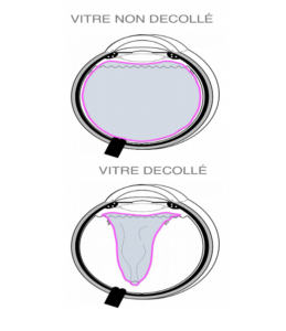
A posterior vitreous detachment occurs when the vitreous envelope (the hyaloid) detaches from the retina to which it normally adheres.
What causes posterior vitreous detachment?
Posterior vitreous detachment is a natural and normal phenomenon that occurs as a result of aging. With time, the vitreous gel that fills the posterior segment of the globe liquefies.
At the same time, its envelope breaks up and detaches from the retina.
The occurrence of posterior vitreous detachment can be favoured by various precipitating factors:
-Strong myopia: the strong myopic eye being longer than the normal eye, the vitreous detaches earlier in life.
the vitreous detaches earlier in life,
-Trauma to the eyeball: any ocular shock can cause vitreous detachment,
-Ocular surgery: any intraocular intervention can modify the position of the internal structures of the eye and induce vitreous detachment.
Ocular surgery: any intraocular procedure can modify the position of the internal structures of the eye and induce vitreous detachment.
What are the symptoms of posterior vitreous detachment?
Once detached, fragments of the vitreous envelope that float in the posterior cavity of the eye may appear in the field of vision as small moving spots with the
eye movements that cause floaters. These vitreous floaters can be perceived as flies, spider webs, commas, filaments… (these are called myodesopsias)
Flashes of light called phosphenes may accompany the floating bodies. These flashes are linked to the traction exerted by the vitreous envelope when it detaches from the retina.
How is posterior vitreous detachment diagnosed?
The diagnosis is made by examining the eye fundus, which shows fragments of
The diagnosis is made by fundus examination, which shows fragments of detached hyaloid floating in the vitreous cavity, mobile with the movements of the globe.
In some cases, a complementary examination by OCT or ultrasound may be useful to look for the persistence of residual pathological adhesions between the retina and the vitreous.
What is the treatment for posterior vitreous detachment?
There is usually no treatment for posterior vitreous detachment. Posterior vitreous detachment
posterior vitreous detachment does not have a potentially deleterious impact on visual function
function except in cases where it is accompanied by the creation of retinal tears.
When detaching from the retina, the release of traction between the vitreous envelope and the retina can cause retinal tears.
A complete examination of the retina will therefore be carried out during the fundus examination to be sure that the detachment of the gel has not created a tear. If a tear is present, treatment with laser photocoagulation will be carried out in order to prevent the occurrence of a
retinal detachment.
In the absence of retinal tears, the evolution of posterior vitreous detachment is marked by the progressive reduction over time (often over several months) of the discomfort caused by the floating bodies (the vitreous envelope progressively settling in the sloping part of the eye and no longer blocking the visual axis).
In exceptional cases, the floaters may remain very annoying. In rare cases, pathological vitreoretinal adhesions may persist (incomplete vitreous detachment).
In such cases, surgical treatment by vitrectomy may be indicated.
After the initial fundus examination is performed rapidly to confirm the diagnosis and rule out the presence of a retinal tear, it may be necessary to repeat the fundus examination at a later stage to rule out the appearance of a tear in the meantime. This is because posterior vitreous detachment is a progressive phenomenon and it is only after some time that the gel envelope is completely detached from the retina. Retinal tears can therefore occur during this time. A sudden change in symptoms (in particular an acute increase in the number of floaters or the occurrence of flashes of light) should lead to a check of the fundus. In the event of constant amputation of the visual field (appearance of a fixed black zone similar to a curtain obstructing part of the field of vision) a new consultation in emergency is necessary (this observation being suggestive of retinal detachment, requiring urgent surgical treatment).
Hoping to have given you a better understanding of the causes and consequences of this condition, we are at your
and consequences of this condition, we are at your disposal for any further information.
CONTACT
You want to make an appointment? You can either call one of the 5 sites via the button below, or make an appointment directly online in the Contact section, or send me a message in the Contact section
The ophthalmology secretaries and Dr. Qin’s team are available to answer all your questions and requests for information in order to make the best choice. I will be pleased to welcome you at one of the 5 sites.